The Ultimate Guide to Camping: Everything You Need to Know
Keep Reading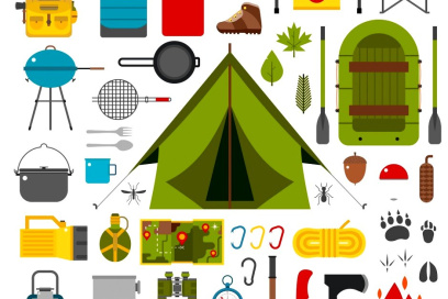
Latest posts
Exploring the Unexpected Link: What Does Camping Mean in Relation to Abortion?
Welcome, adventurers! Welcome to my camping blog where the boundaries of expectation will be broken and where the mysterious worlds of camping and abortion will collide in an unbelievable tapestry...
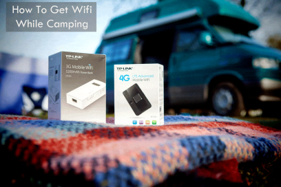
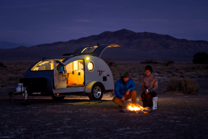
Stay Connected in the Great Outdoors: How to Get WiFi While Camping
Experience the magic and wonder of camping, where nature blends harmoniously with modern connectivity. Today's digital landscape makes staying connected a necessity, even in remote locales where co...
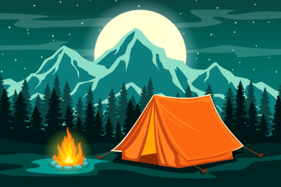
Trailblazing Through the Tent Jungle: Picking Your Ideal Camping Companion
Are You Prepared for an Enthralling Odyssey in Search of the Ideal Camping Tent? Fear Not - we shall untie all its hidden myster...
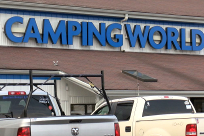
Unlock the Doors: Is Camping World Open Today? Your Complete Guide!
Hello outdoor enthusiasts!
Budgeting for Adventure: Understanding the True Costs of Camping
Prepare yourself for an eye-opening adventure into the mysterious depths of camping costs! Venture forth...
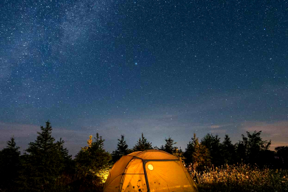
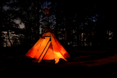
From Campouts to Camp Outs: The Ultimate Guide to Nature's Best Adventure
Prepare yourself for an extraordinary adventure, my friend. Imagine leaving behind the hustle and bustle of everyday life behind to plunge headfirst into Mother Nature's beauty and splendor. Begin...
About Us
Welcome on GlobeHealthTours, our passionate outdoor blog. With years of experience exploring nature's splendor, we have become experts at everything camping related - from choosing an appropriate tent to crafting delicious campfire meals! Through this blog, we share our expertise to inspire other to experience its joys first-hand - join us as our journey across our great outdoor to uncover top camping locations and create lifelong memories in nature!